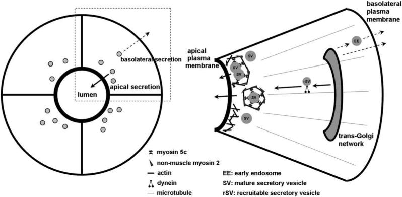
Scheme 1.

The diagram to the left depicts individual lacrimal acinar cells organized around a central lumenal region, as well as the directions for apical and basolateral secretion of proteins from the cell. The apical plasma membrane domain can be distinguished in intact tissue and in the in vitro reconstituted cultures by the intense labeling associated with the subapical actin filament network. The diagram to the right depicts a single acinar cell expanded from the panel to the left. SV are shown concentrated in the subapical cytoplasm beneath the actin-enriched apical plasma membrane. Some SV are undergoing fusion, by being enveloped in actin coats enriched in non-muscle Myo2 and Myo5c, adjacent to spots where actin filaments have transiently disassembled beneath the apical plasma membrane. Exocytosis using these mechanisms is elicited by secretagogue stimulation (bold arrows). rSV are also shown moving towards the apical plasma membrane to sustain the secretory response, a response evoked by secretagogue stimulation (bold arrows). Other vesicular traffic emerging from the trans-Golgi network can be sorted to the basolateral membrane for constitutive exocytosis (dashed arrows).
