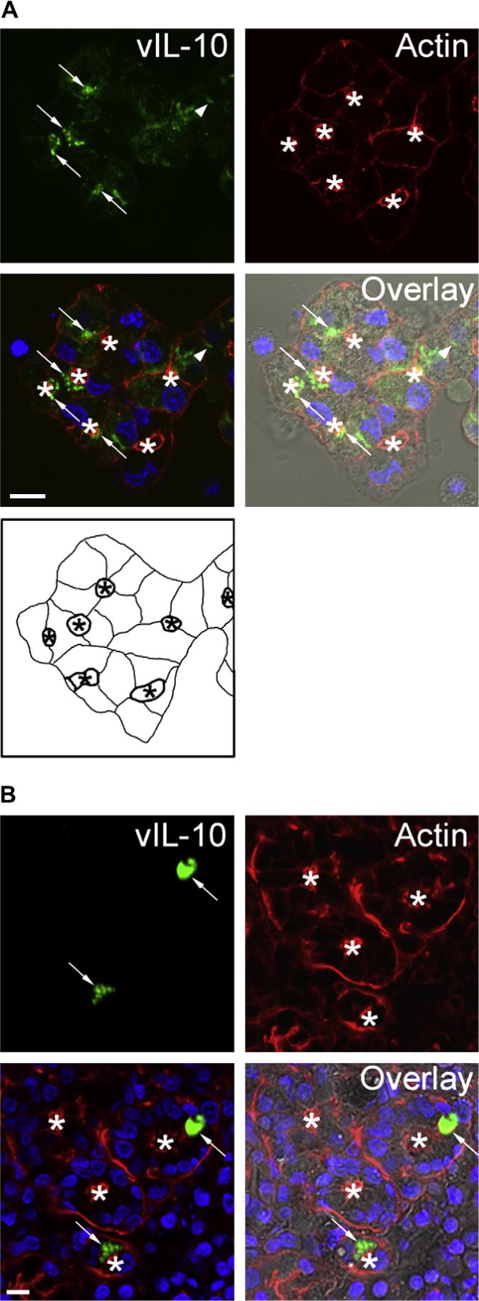
Fig. 1.

vIL-10 immunoreactivity is detected in large intracellular vesicles in transduced LGAC. A. Rabbit LGACs were seeded onto Matrigel-coated 18 mm circular glass coverslips in 12-well plates at 2 × 10⁶ cells/well. On day 2 of culture, reconstituted acini were transduced with AdvIL-10 at a MOI of 5 for 24 h. Cells were then fixed and processed to fluorescently label vIL-10 (green), actin filaments (red) and cellular nuclei (blue). The DIC image is also shown for comparison. Arrows, accumulation of vIL-10 in large vesicles beneath the actin-enriched apical plasma membrane; arrowhead, accumulation of vIL-10 in basolateral structures; bar, 10 μm and *, apical/lumenal region. A schematic diagram of the acini transduced with AdvIL-10 is also shown for reference. The lumen (indicated with an asterisk) is bounded by the apical plasma membranes (thick lines). Basolateral plasma membranes are illustrated as thinner lines. B. Immunocytochemistry studies of IL-10 expression at 5 days post-injection in rabbit inferior lacrimal glands. AdvIL-10 (1 × 10⁸ PFU in 0.2 mL) was injected into each rabbit's inferior lacrimal gland as described in Section 2. 5 days after inoculation, rabbits were sacrificed and inferior lacrimal glands were removed from each animal. After fixing in 4% paraformaldehyde and sucrose rehydration, the glands were embedded in OCT and frozen sections were processed to fluorescently label vIL-10 (green), actin filaments (red) and cellular nuclei (blue). The DIC image is also shown for comparison. Arrows, accumulation of vIL-10 in vesicle-like structures beneath the apical plasma membrane; bar, 10 μm and *, apical/lumenal region.
