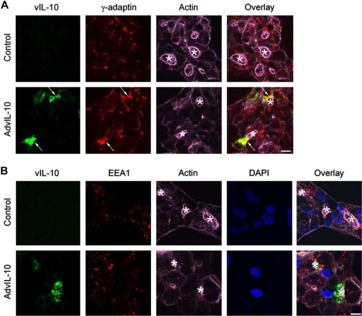
Fig. 2.

vIL-10 is partially co-localized with biosynthetic but not basolateral endosomal markers. Rabbit LGACs were cultured and transduced with AdvIL-10 as described in legend for Fig. 1A. Cells were then fixed and processed to fluorescently label vIL-10 (green), γ-adaptin (A) or EEA1 (B) (red), actin filaments (purple) and DAPI (blue). Arrows, co-localization of vIL-10 and γ-adaptin; bar, 10 μm and *, apical/lumenal region.
