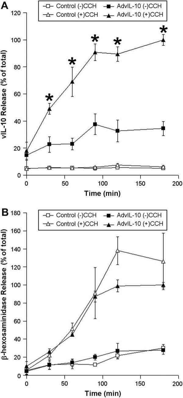
Fig. 4.

Time course of CCH-stimulated release of vIL-10 and β-hexosaminidase from LGAC. Rabbit LGACs were cultured and transduced with AdvIL-10 as described in the legend for Fig. 3. After treatment with or without 100 μM CCH at time intervals from 0 to 180 min, aliquots of the medium were collected and processed for measurement of vIL-10 (A) and β-hexosaminidase (B) release using ELISA and biochemical assays, respectively. vIL-10 and β-hexosaminidase release are plotted as % of total, with total defined as the maximal release elicited by 100 μM CCH at 180 min. n = 3 and *, significant at p ≤ 0.05, AdvIL-10 (+)CCH versus AdvIL-10 (−)CCH. Error bars represent s.e.m.
