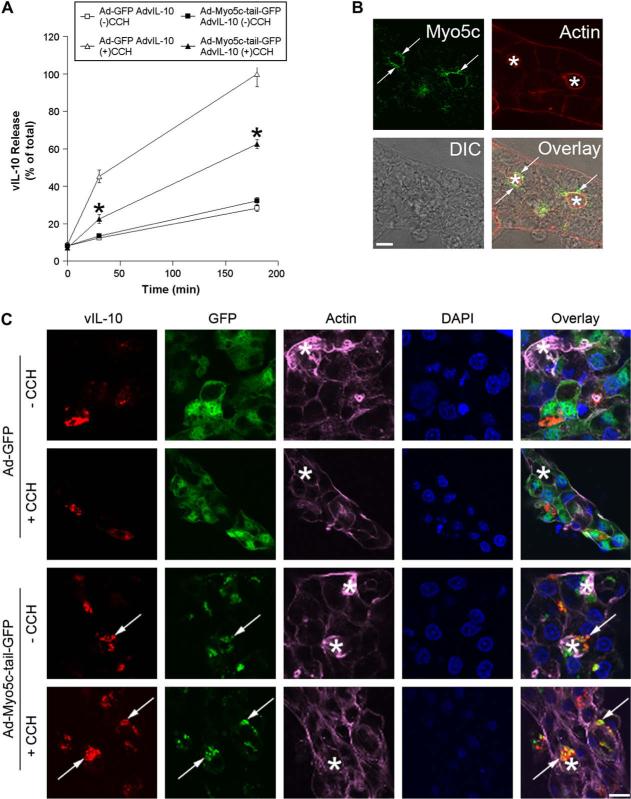
Fig. 8.
Myosin 5c participates in vIL-10 secretion from LGAC. A. Effect of Ad-Myo5c-tail-GFP transduction on CCH-stimulated release of vIL-10 from LGAC. Rabbit LGACs were seeded onto Matrigel-coated 24-well plates at 1 × 10⁶ cells/well. On day 2 of culture, reconstituted acini were singly or doubly-transduced with Ad-GFP, Ad-Myo5c-tail-GFP or AdvIL-10 at a MOI of 5 for 24 h. Control and transduced acini were then incubated in fresh medium for 1 h to allow cell equilibration. After treatment with or without 100 μM CCH at time intervals from 0 to 180 min, aliquots of the medium were collected and processed for measurement of vIL-10 release using ELISA assays. vIL-10 release is plotted as % of total, with total defined as the maximal release elicited by 100 μM CCH at 180 min in acini doubly-transduced with AdvIL-10 and Ad-GFP. n = 10 and *, significant at p ≤ 0.05, Ad-Myo5c-tail-GFP AdvIL-10 (+)CCH versus Ad-GFP AdvIL-10 (+)CCH. Error bars represent s.e.m. B. Rabbit LGACs were seeded onto Matrigel-coated 18 mm circular glass coverslips in 12-well plates at 2 × 10⁶ cells/well. On day 3 of culture, reconstituted acini were fixed and processed to fluorescently label Myo5c (green) and actin filaments (red). The DIC image is shown for comparison and the overlay image shows the super-imposition of the fluorescence image and the DIC image. Arrows, large apparent SV enriched in Myo5c; bar, 5 μm and *, apical/lumenal region. C. Effect of Ad-Myo5c-tail-GFP transduction on vIL-10 distribution in LGAC. Rabbit LGACs were seeded onto Matrigel-coated 12-well plates at 2 × 10⁶ cells/well. On day 2 of culture, reconstituted acini were singly or doubly-transduced with Ad-GFP, Ad-Myo5c-tail-GFP or AdvIL-10 at a MOI of 5 for 24 h. Cells were then fixed and processed to fluorescently label vIL-10 (red), GFP (green), actin filaments (purple) and cellular nuclei (blue). Arrows, co-localization of vIL-10 and Myo5c-tail-GFP; bar, 10 μm and *, apical/lumenal region.

