Fig. 3.
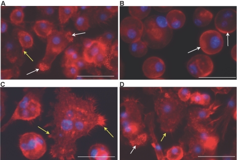
MMP-9 null macrophages display delayed and abnormal fusion-induced cytoskeletal reorganization. Representative images of WT (A and C) and MMP-9 null (B and D) IL-4-treated macrophages from Day 1 (A and B) and Day 3 (C and D) culture stained with phalloidin and DAPI are shown. At Day 1, WT macrophages exhibited normal elongation, accumulation of punctate actin (white arrows), and lamellipodia formation (yellow arrows), which were absent in MMP-9 null macrophages. Arrows in B identify the presence of peripheral actin. At Day 3, WT macrophages displayed FBGC formation and extensive lamellipodia formation (yellow arrows in C). (D) At this time-point, MMP-9 null macrophages displayed changes consistent with preparation for fusion, such as lamellipodia formation (yellow arrow) and cytoskeletal reorganization (white arrow), but FBGC formation was not evident. Original bars = 25 μm (A–D).

