
Figure 1.
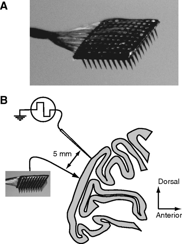
Experimental methods. A, A photograph of the array. The 10 × 10 grid of silicone microelectrodes had a 400 μm spacing, 1.0 mm length, and was inserted 0.6 mm into cortex. B, A diagram of the recording arrangement on the operculum of V1, shown in a sagittal section of macaque cortex. In two experiments, we simultaneously recorded from a group of seven linearly arranged microelectrodes, separated by at least 5 mm from the array.