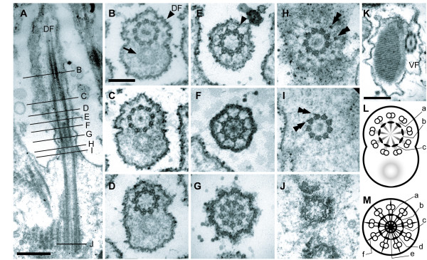
Figure 6.
Transmission electron micrographs (TEM) showing paraxonemal rods in the flagella, the flagellar transition zone and the basal bodies of Calkinsia aureus. A. Longitudinal section of the dorsal flagellum (DF) showing the flagellar transition zone and the dorsal basal body (DB) (bar = 500 nm). B-J. Non-consecutive serial sections through the DF (B), the flagellar transition zone (C-G), and the DB (H-J) as viewed from anterior end (images at same scale, bar = 200 nm). B. Section showing the 9+2 configuration of axonemal microtubules and the tubular paraxonemal rod (arrow) in the DF. C. Section showing termination of central microtubules and the 9+0 configuration of axonemal microtubules. D. Section showing the transition zone through an outer concentric ring associated with nine electron dense globules inside of each doublet and faint spokes that extend inward from the each globule (see L for a diagram of this micrograph). E. Section through the nine radial connectives (arrowhead) that extend outward from each doublet to the flagellar membrane. F. Section showing the radial connectives that extend outward toward the flagellar membrane, the spokes that extend inward from the microtubular doublets, the central electron dense hub, and inner concentric rings (see M for the diagram of this micrograph). G. Section showing the electron dense hub and inner and outer concentric rings, and the absence of radial connectives. H. A section at the level of the insertion of the DF. The transitional fibers (double arrowheads) extending from the microtubular triplets of the DB are shown. I. Section through the area just below the distal boundary of the DB. The transitional fibers (double arrowheads) connect to each microtubular triplet. J. Section through the proximal region of the DB showing the cartwheel structure. K. View through the paraxonemal rod of the ventral flagellum (VF) (bar = 500 nm). L. Diagram of the level of D showing faint spokes (a) that extend inward from each globule, an outer concentric ring (b) and nine electron dense globules (c). M. Diagram of the level of F showing spokes (a), an outer concentric ring (b), nine electron dense globules (c), an electron dense hub (d), an inner concentric ring (e) and radial connectives (f).

