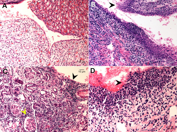
Figure 3.
Summary of the kidney lesions found in F344 rats experimentally infected with U. parvum. Panel A is a 200× magnification of kidney tissue from a Control rat and demonstrates the lack of inflammatory lesions that are characteristic in animals inoculated with U. parvum. Panels B, C, and D are tissue sections from animals inoculated with U. parvum that had a lesion score of 4 for total area affected. Panel B is a 400× magnification demonstrating the inflammatory infiltrate extending from the renal pelvic space into the interstitium with uroepithelium largely intact. The black arrow is pointing to uroepithelial hyperplasia. Panel C is a 400× magnification of renal tubules. The black arrow is pointing to the extensive inflammatory infiltrate throughout the renal tubular interstitium. The yellow arrow is pointing to a glomerulus. Panel D is a 600× magnification of renal uroepithelium at the edge of the pelvic space. The black arrow is pointing to extensive hemorrhage and disruption of the uroepithelial barrier by a fibrinous inflammatory infiltrate.

