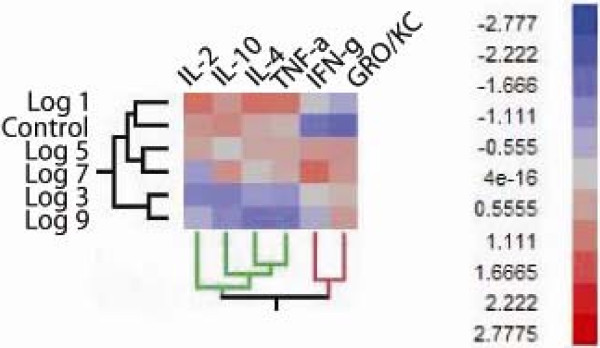
Figure 7.
Profiling the inflammatory response to different doses of U. parvum in culture negative F344 rats. Panel A is a clustered heat map representing the standardized LS means for each cytokine with a significantly different pattern of expression among infection dose groups (P 0.05). Values were obtained by one-way ANOVA using a row by row modeling with Fischer C correction for multiple comparisons. Two main cytokine cluster patterns were identified in the analysis and are demarcated by the green and red cluster tree. The number of biological replicates were n = 6 for control, n = 10 for log 1 CFU, n = 11 for log 3 CFU, n = 8 for log 5 CFU, n = 5 for log 7 CFU, and n = 3 for log 9 CFU. The red arrow is highlighting the pattern of cytokines present in the urine of culture negative rats that were inoculated with log 5 CFU. Animals within this dose group were the only animals to exhibit an obvious inflammatory cell infiltrate comprising a mixture of mononuclear cells with neutrophils (P 0.006, Figure 2, Panel D).

