
Figure 2.
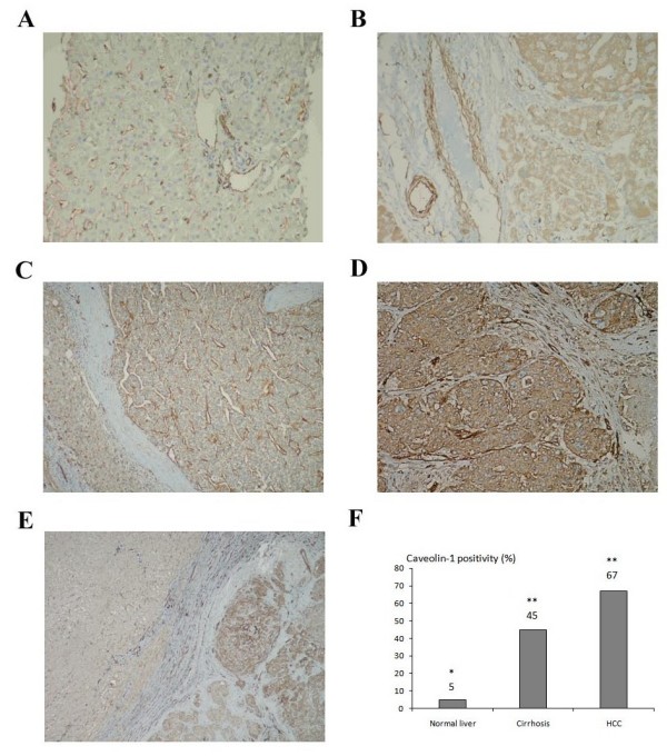
Immunohistochemical staining of Cav-1 in normal, cirrhotic and HCC tissue sections. (A) Absence of Cav-1 expression in hepatocytes of normal donor liver tissue and strong Cav-1 expression in the endothelium of the blood vessels. (B) Cav-1 positivity in the cirrhotic nodule and endothelium of the vessels with same intensity. (C) Membranous Cav-1 positivity in one of the well differentiated HCC cases. (D) Strong cytoplasmic Cav-1 expression in a poorly differentiated HCC. (E) Staining of Cav-1 in an HCC sample with its tumoral and surrounding peritumoral region. (F) The percentage of Cav-1 staining in normal liver, cirrhosis and HCC tissues (* p < 0.001 when HCC and cirrhotic tissues were compared to normal liver; ** p = 0.002 when HCC was compared to cirrhotic tissues). Normal liver tissues, cirrhotic tissues (without HCC) and HCC tissues are taken from different patients.