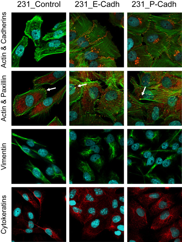
Figure 2.
Expression of E- or P-cadherin does not completely reverse the mesenchymal phenotype of 231 cells. First row: Double immunofluorescence of E- or P-cadherin (Alexa-594, red) and actin cytoskeleton (Phalloidin-Alexa-488, green). Second row: Focal adhesions (arrows) co-stained with Phalloidin (green) and anti-paxillin (red). Note that no evident changes in the number or organization of focal adhesions are seen among the different conditions. Third and fourth rows: expression of the mesenchymal marker vimentin and the epithelial marker cytokeratin.

