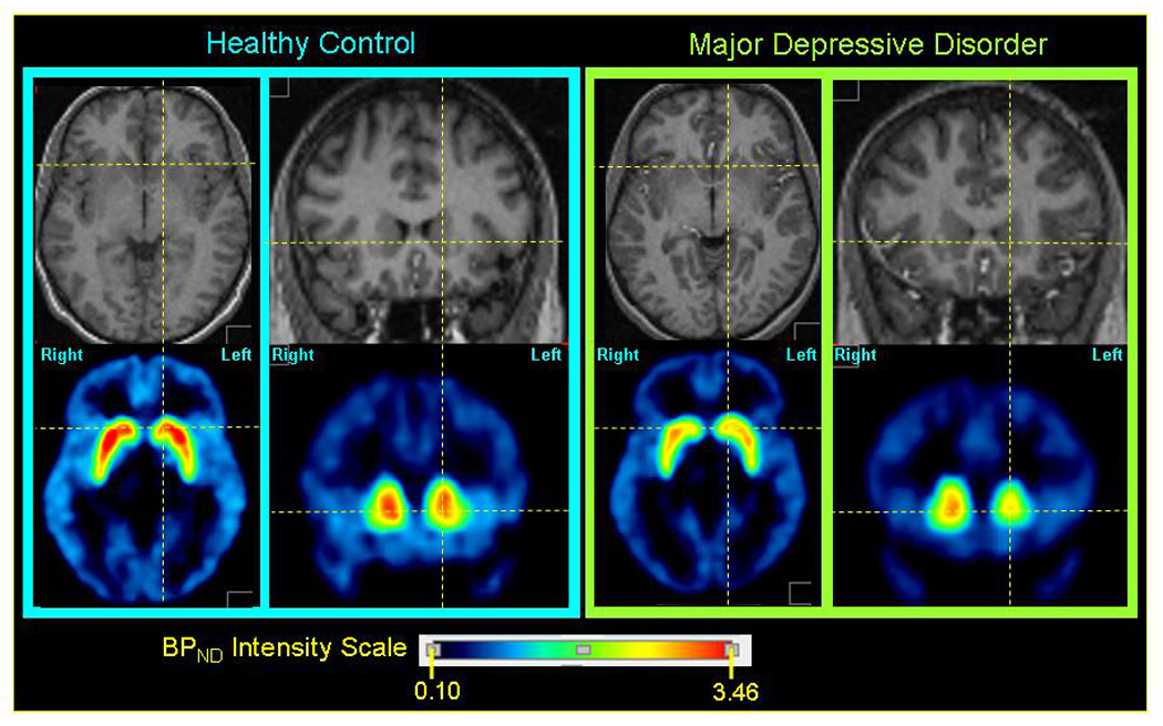
Figure 2. Representative parametric dopamine-D1 receptor BPND images through the striatum in a control subject (left panel) and an MDD subject (right panel).

Within each panel the upper row of images show axial (left) and coronal (right) sections through the anatomical MRI, on which the regions-of-interest (ROI) were defined (Drevets et al. 2001), and the lower row of images show parametric images of the non-displaceable component of the D1-recetor binding potential (BPND) modeled from PET emission images. The MRI and PET images for each subject are co-registered. The axial images show the plane containing both the anterior and posterior commissures. The orthogonal lines locate approximately the center of the caudate head on this bi-commissural plane. The coronal sections pass through the anterior striatum, which at the level shown is composed predominantly of the caudate head. The middle caudate region is evident in the coronal sections, where it is situated immediately above the bicommissural plane (marked by the horizontal line). The D1-receptor BPND consistently was higher in the right caudate than the left in both groups (see figure 1, figure 3), as evinced in these representative images. The mean BPND value in the left middle caudate of the MDD group was lower than in the control group (figure 1).
