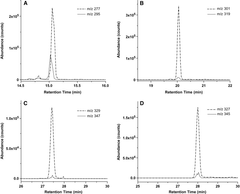
Fig. 1.
Selected ion chromatograms of major 13C-labeled n-3 PUFA isotopomers versus unlabeled endogenous components in plasma at the end of U-[13C]α-linolenic acid ([13C]α-LNA) infusion. The m/z value of each ion indicated was used to detect the M-pentafluorobenzyl ([M-PFB]−) ions of each unlabeled and stable labeled n-3 PUFA, respectively. Depicted are the various isotopomers of (A) α-linolenic acid (α-LNA) (18:3 n-3), (B) eicosapentaenoic acid (EPA) (20:5 n-3), (C) docosapentaenoic acid (DPA) (22:5 n-3), and (D) docosahexaenoic acid (DHA) (22:6 n-3).

