
Figure 3. Defective oligodendrocyte differentiation in yy1 conditional-knockout (cko) mice is associated with persistent global histone acetylation.
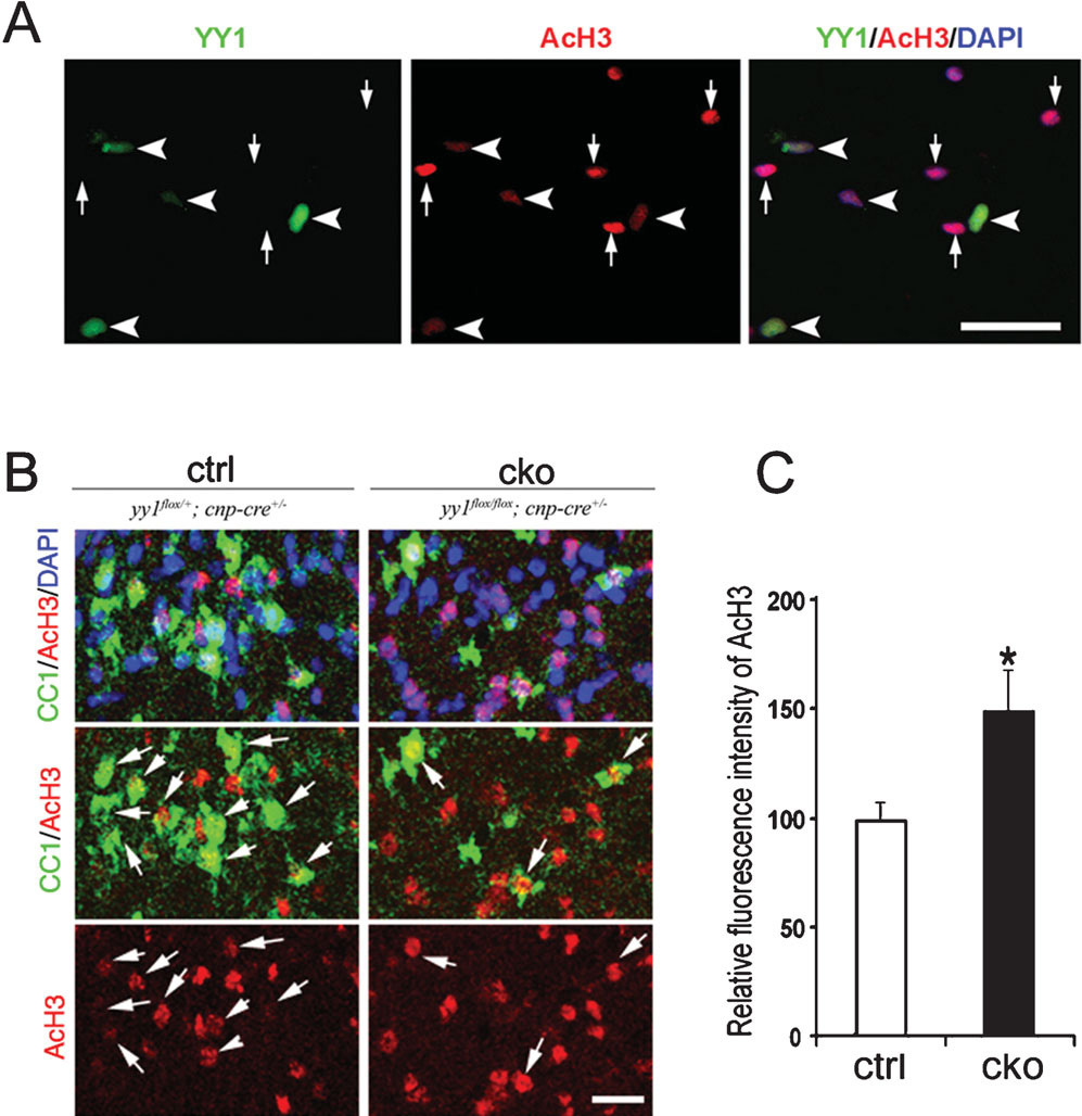
(A) Oligodendrocyte progenitors generated from neonatal yy1flox/flox mice were infected with adenovirus-CMV-Cre and 48-hours later stained for YY1 (green) and AcH3 (red). Note that YY1-deleted cells (arrowhead) have higher AcH3 immunoreactivity compared to uninfected cells (arrow) in the same field. Scale bar, 20 µm. (B) Sagittal sections of the cerebellum of control and yy1-cko mice stained for oligodendrocyte marker CC1 (green) AcH3 (red) shows a higher level of AcH3 in yy1-cko mice compared to sibling controls at P18. Scale bar, 20 µm. (C) Fluorescence intensity of AcH3 in CC1+ cells in control mice (n = 3) and yy1-cko mice (n = 3) was calculated using NIH ImageJ on acquired confocal images. The average fluorescence intensity in the control siblings is set arbitrarily at 100. Data are mean ± SD. *P<0.05, Student’s t-test.