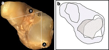
Fig. 2.

a Inferior view of left clubfoot talus (grade IV). Angle β formed by a longitudinal axis A of the posterior facet for the calcaneus with a line B parallel to the anterior trochlea border. b Analogue drawing. The joint surface of a normal foot is speckled grey, the one of a clubfoot white
