
Fig. 6.
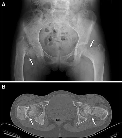
Use of CT imaging in FD. a This radiograph of a 10-year-old girl with FD revealed severe disease in both femurs, with a suggestion of a fracture. At the time of the radiograph, she was asymptomatic and ambulating without assistance. b CT at the level of the femora neck revealed the true extent of the disease, with almost complete disruption of the neck (arrows). The extent of the disease was not fully appreciated on the radiograph