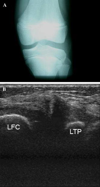
Fig. 1.

a Plain X-ray (anterior–posterior view) of a 10-year-old boy’s knee demonstrating widening of the lateral joint space, squaring of the lateral femoral condyle, cupping of the lateral tibial plateau, and tibial eminence hypoplasia. b An ultrasound of the wide and irregular lateral meniscus (LFC lateral femoral condyle, LTP lateral tibial plateau)
