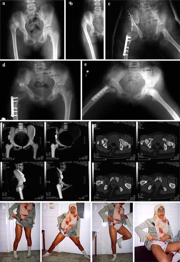
Fig. 3a–k.

Right developmental dysplasia of the hip (DDH) in an 11-year-old girl with excellent outcome. a, b Pre-operative radiograph in internal rotation revealing a normal neck shaft angle with a relatively small femoral head. c Post-operative reduction, well-contained head, triple pelvic osteotomy with the excised femoral segment used as a graft to maintain acetabular displacement. d, e Radiograph at 2 years follow up in full abduction with extended hips. f, g Multislice computed tomography (CT) sections and 3D reconstruction revealing normal anteversion angle and a normal hip articulation. h–k Photographs of the patient at 17 years of age with a mobile hip performing all movements
