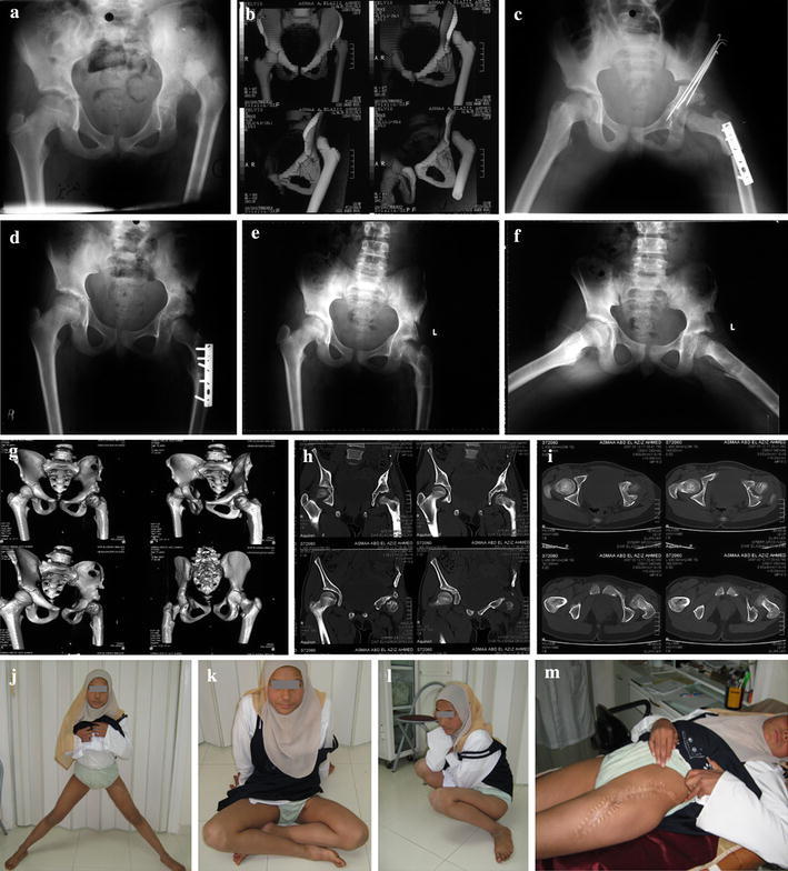
Fig. 4a–m.

Left DDH in a 12-year-old girl with excellent outcome. a, b Pre-operative radiograph and 3D reconstruction CT views showing a highly dislocated relatively small femoral head. c Post-operative radiograph, with well-contained femoral head following 5 cm femoral shortening with derotation, minimal varization Salter pelvic osteotomy. d Two years follow up radiographs. e, f Three years follow up radiographs with full abduction in extension. g–i Three years follow up CT with reconstruction views showing well-contained femoral head with normal anteversion angle. j–m Photographs showing full range of movements of the left hip, yet with an unsightly scar
