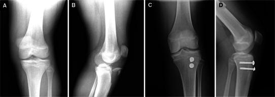
Fig. 2.

a Plain radiograph AP view of a 14-year-old with type IIIB tibial tuberosity fracture. b Lateral radiograph of the same patient demonstrating a fragment comminution. c Six-month post-op AP radiograph of the same patient demonstrating fixation. d Lateral radiograph demonstrating union with good remodeling
