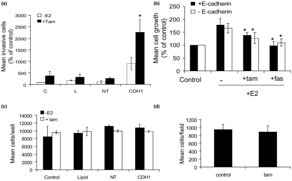
Figure 3.
Tamoxifen promotes invasion of E-cadherin deficient MCF-7 cells. (a) MCF-7 cells were left untreated (control, 'C') or treated with transfection lipid ('L'), non-targeting (scrambled) siRNA ('NT') or E-cadherin-specific siRNA ('CDH1') for 72 hours before assessing their invasive capacity in oestrogen-free and tamoxifen-containing medium. siRNA-mediated suppression of E-cadherin expression alone promoted an increase in cell invasion through Matrigel. Inclusion of tamoxifen in CDH1-treated cells resulted in a further increase in invasive capacity. The graph is the mean of three separate experiments. *p < 0.01 versus CDH siRNA treatment. (b) MCF-7 proliferation was determined in response to E2 ± tamoxifen and fulvestrant in the presence and absence of E-cadherin. Data demonstrated that these cells were responsive to both E2 and anti-oestrogens irrespective of E-cadherin presence. *p < 0.05 versus E2 alone. (c) The growth of untreated MCF7 cells or cells treated with lipid (L), non-targeting siRNA (NT) or CDH-1 siRNA (CDH1) over five days was determined by coulter counting. No treatment significantly affected cellular growth over this time period. (d) MDA-MB-231 cells were treated with siRNA ± tamoxifen and changes in their invasion determined. Tamoxifen did not have any significant effect on MDA-MB-231 cell invasion.

