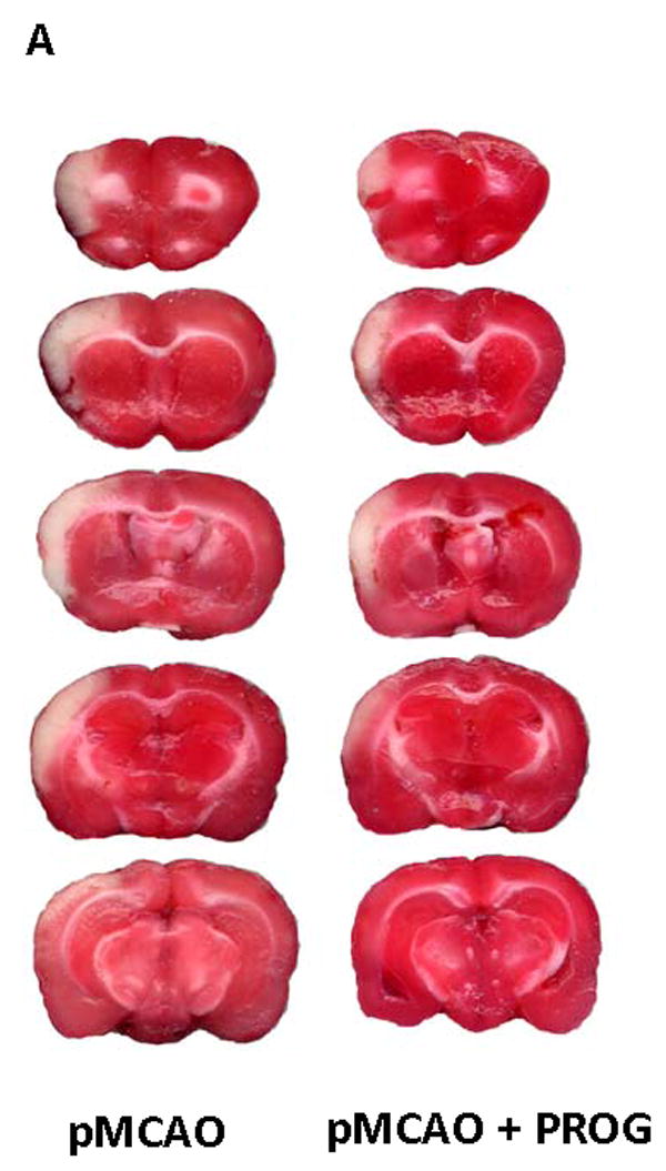
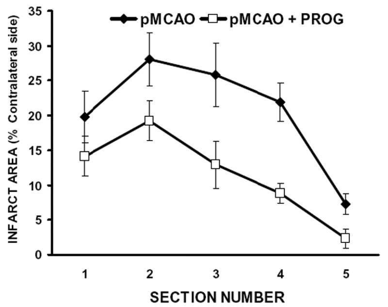
Figure 1. PROG reduces infarct volume in a rat model of pMCAO.


A) TTC-stained coronal sections from representative rats given either vehicle or PROG, brains harvested at 72h post occlusion. Infarcts are shown as pale (unstained) regions. The infarct area in PROG-treated rats is substantially reduced. B) Line graph shows the percent area distribution of infarction to the area of the contralateral side in each of five forebrain sections in pMCAO and pMCAO plus PROG treated groups. Values are mean ± SD.
