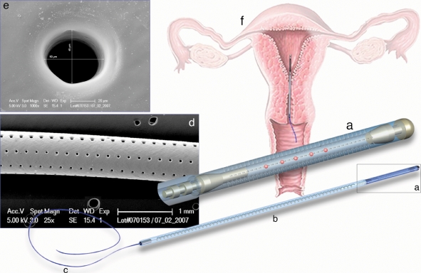
Figure 1.
Diagram of the IUCS used in women undergoing ICSI: (a) microdrilled silicone segment containing the oocytes/embryos; (b) stabilization segment; (c) extraction segment; (d) scanning electron microscopy of the drilled silicone segment (magnification ×25); (e) high magnification of a laser-drilled hole (scanning electron microscopy, magnification ×1000); (f) schematic representation of the device position in the uterine cavity.

