
Fig. 1.
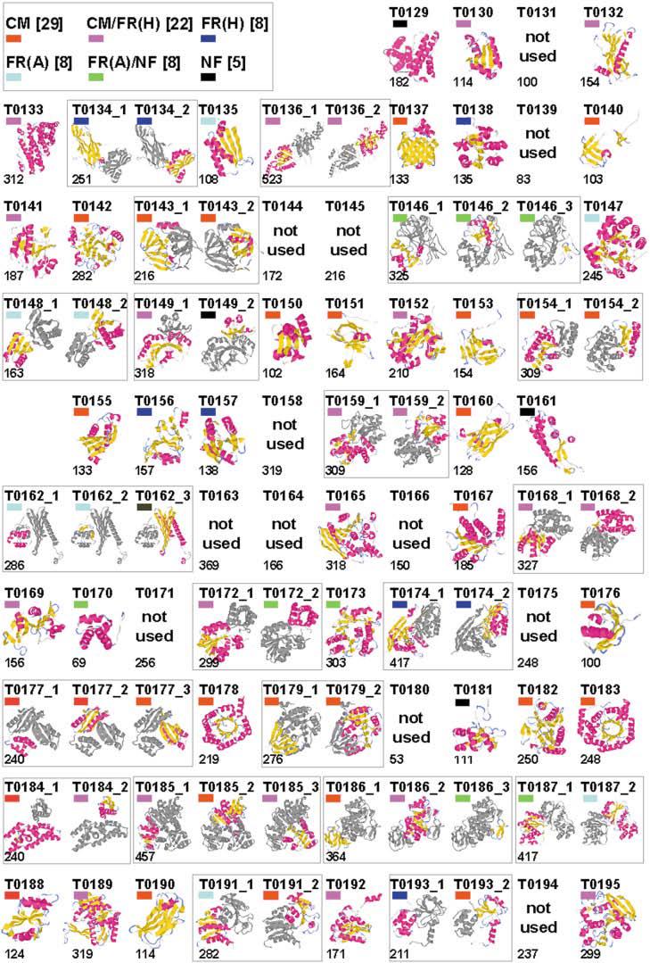
CASP5 domains. Thumbnail images of CASP5 targets, created by using the graphics program RasMol.30 Models of the CASP5 target structures are split into domains, and indicated domains are colored according to secondary structural elements: α-helix (pink) and β-sheet (yellow), with the remaining secondary structural elements colored gray. The number to the lower left of each image indicates the target length, and colored block to the upper left of each image indicates the target classification: CM (red), CM/FR(H) (pink), FR(H) (blue), FR(A) (cyan), FR(A)/NF (green), and NF (black).