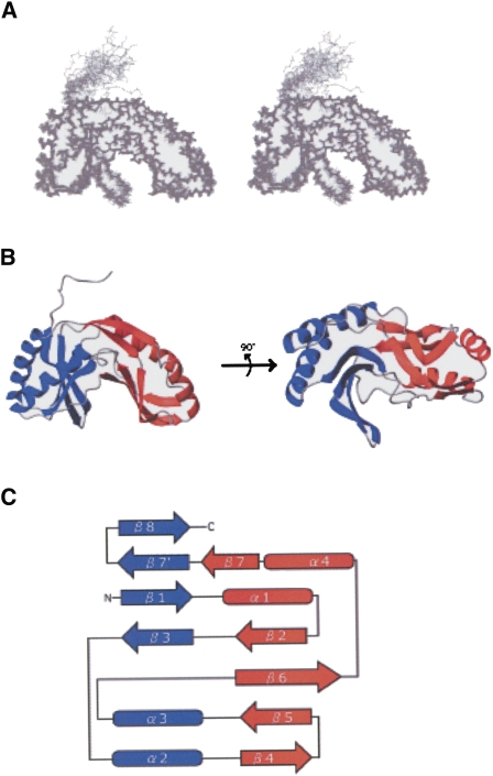
FIGURE 6.
Tertiary structure of PF0027 protein. (A) Stereo view of superposition of final 20 structures obtained by NMR structure determination. (B) Ribbon diagram. (Blue) The terminal lobe; (red) the transit lobe. (C) Schematic diagram of secondary structure topology. (Arrows) Secondary structure elements (β-strands) and cylinders (α-helices).

