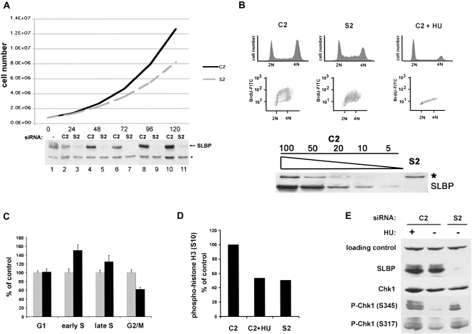
FIGURE 1.
SLBP knockdown cells are viable but grow at a slower rate as a result of various S-phase defects. (A) Growth curve (total cell number) of control (C2) and SLBP (S2) RNAi populations. Lysates from the time points shown in the graph were probed for levels of SLBP by Western blotting; (*) cross-reacting band. (B) SLBP knockdown results in S-phase accumulation and a reduced rate of DNA replication. C2 and S2 treated (72 h) U2OS cells were pulsed for 1 h with 10 μM bromodeoxyuridine (BrdU) and harvested and analyzed by FACS for DNA content and DNA synthesis. Hydroxyurea (HU; 5 mM) was added to a parallel culture of C2 cells 1 h prior to the addition of BrdU. Cells were fixed and BrdU incorporation detected with an anti-BrdU antibody conjugated to FITC and for DNA content with propidium iodide. Cells were analyzed using a FACScan device and Summit software for DNA content and intensity of BrdU incorporation. The levels of SLBP in a typical experiment are shown below the graph. They were determined by Western blotting compared to a dilution series of a control extract (1:2 to 1:20). (C) The cell cycle distribution (G1, early and late S phase, and G2/M) was quantified from the data in B ([gray bars] C2; [black bars] S2) from three independent experiments. Quantities are expressed as a percentage of the C2 control cells. (D) Parallel cultures from B were fixed and stained with an antibody to the mitotic marker phospho-histone H3, and the antibody was detected with a secondary antibody conjugated to FITC. Cells were analyzed similarly as in B, and the percentage of cells with fluorescent signal in the G2 population was quantified. The number of phospho-histone positive cells is expressed as a percentage of the C2 control cells. (E) SLBP knockdown results in the activation of a Chk1-dependent cell cycle checkpoint. Protein lysates from SLBP knockdown cells were analyzed by Western blotting for SLBP, total Chk1 protein, and two phosphorylation sites (serines 317 and 345) using phospho-specific Chk1antibodies. Cells treated with HU for 1 h were used a positive control for Chk1 activation.

