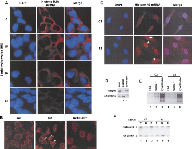
FIGURE 4.
Histone mRNA in SLBP knockdown cells is retained in the nucleus. (A) HeLa cells synchronized by double-thymidine block were released into S phase for 3 h and treated with 5 mM hydroxyurea (HU) for 0, 15, 30, and 45 min. Cells were fixed and prepared for in situ hybridization with antisense DIG-labeled H2a mRNA. Cells were stained with DAPI and anti-DIG antibodies, (blue) DAPI, and (red) histone H2A mRNA. (B) Logarithmically growing HeLa cells were treated with the indicated siRNAs, and in situ hybridization was performed with antisense DIG-labeled H3 mRNA. Note that histone mRNA accumulates in (arrows) nucleoli of HeLa cells in S2-treated cells. (C) U2OS cells were treated with the indicated siRNAs, and in situ hybridization was performed with antisense DIG-labeled H3 mRNA as in B. Cells were stained with DAPI. Note that there is no accumulation of histone mRNA in (arrows) nucleoli in U2OS cells. (D) U2Os cells were fractionated into nuclear and cytoplasm and protein or (E,F) RNA from total and subcellular fractions analyzed. (Lane 1) Total, (lane 2) nuclear, and (lane 3) cytoplasmic protein fractions were separated by 12% polyacrylamide gel electrophoresis and probed for Hsp90 (top, as a cytoplasmic marker) and fibrillarin (bottom, as a nuclear marker by Western blotting. (E) Total (10%) input, nuclear, and cytoplasmic RNA fractions from (lanes 1–3) C2 and (lanes 4–6) S2-treated cells were separated on a 1% agarose gel, and rRNA (as a cytoplasmic marker) was visualized by ethidium bromide staining. (F) Total, nuclear, and cytoplasmic RNAs were analyzed by Northern blot analysis, and histone H3 mRNA and U1 snRNA (nuclear marker) were simultaneously detected using a mixture of radiolabeled probes.

