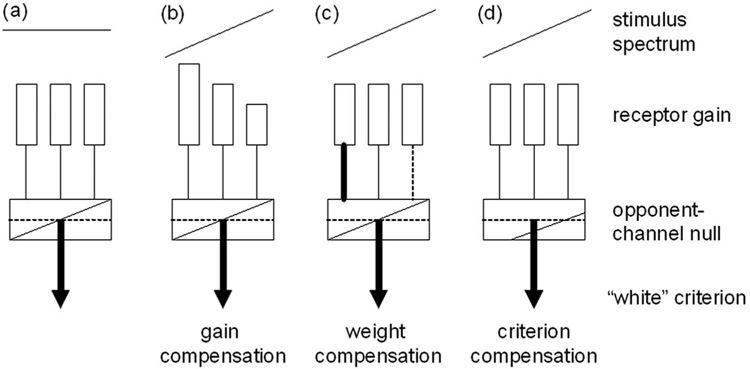
Fig. 3.
Norms in color vision. (a) “White” is assumed to be represented by balanced activity across the S, M, and L cone receptors and by a null response in postreceptoral opponent channels. Responses to a biased spectrum could be renormalized by compensatory adjustments (b) in the sensitivity of the receptors, (c) in the strength of inputs to the opponent channels, or (d) by changing the criterion for white.

