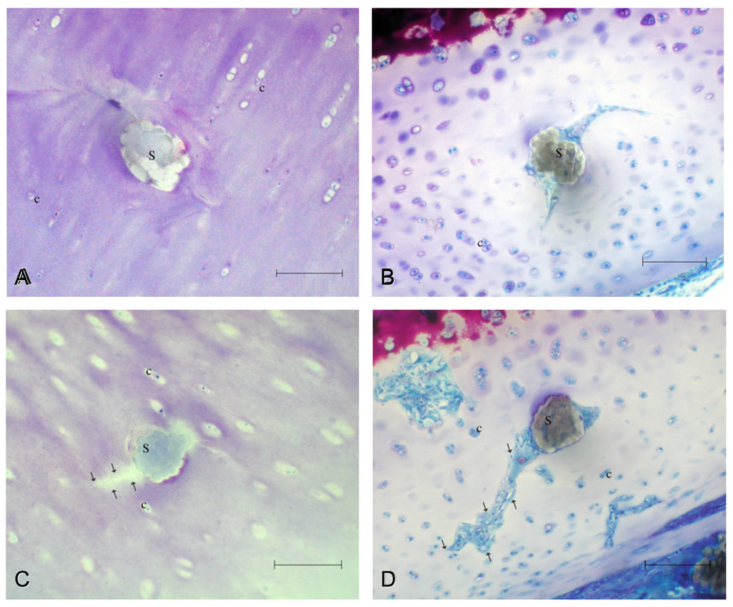
Fig. 4.
Light micrographs of cross-sectioned sutures (S) 2–3 hours after surgery (A, C) and 3 weeks later (B, D). Fissures that developed in the walls of the suture channels [see arrows in (C) and (D)] propagated with time, and at the 3-week juncture, they were filled with a primitive type of avascular scar tissue (D). c = chondrocytes. Bar = 100 µm.

