
Fig. 1.
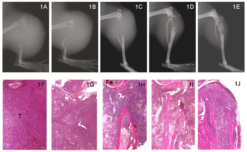
Representative radiographs (top panel; 1A-1E) and H&E histologic sections (bottom panel; 1F-1G) of SCID mice tibias at 6 weeks following intratibial injection of tumor cells. There is no cortical bone present in the proximal tibias of PC-3 (1A & 1F) and PC-3+EV (1B & 1G). However, there is limited destruction of proximal tibias in PC-3+RANK: Fc (1D & 1I) and PC-3 RetroNog + RANK: Fc (1E & 1J) compared to control animals and PC-3 retronog treated tibias (1C & 1H) at 6 weeks. Fe, femur; T, Tumor cells