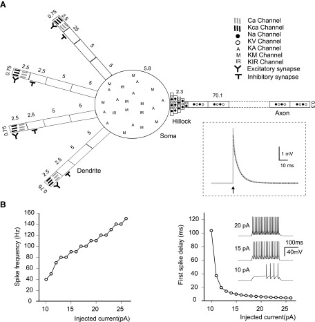
FIG. 5.
Granule cell multi-compartmental model. A: Schematics of the compartmental structure and ion channel distribution in the model (the axon is only partly represented). All compartments are modeled as cylinders (Rall 1968) and their length and diameter (in μm) are indicated. Inset: the voltage transient elicited by a short current pulse (arrow) injected into the soma. Note the biphasic voltage decay (gray trace), from which the time constants τm and τ1 were estimated through bi-exponential fitting (dashed black trace). B: the plots show average firing frequency (left) and 1st-spike latency (right) during injection of depolarizing current steps in the soma. Inset traces: the response of the model to 10-, 15-, and 20-pA current injection.

