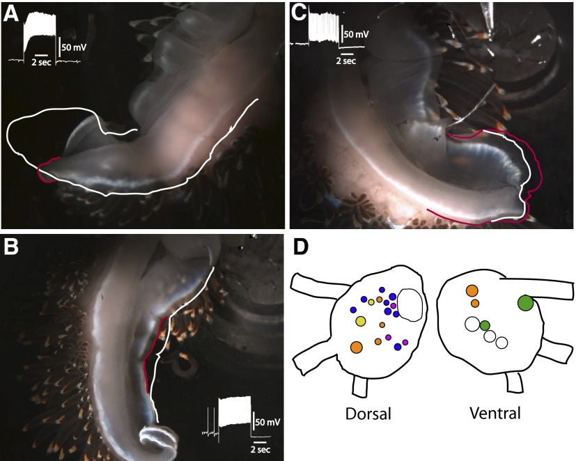
FIG. 1.
Photographs of representative semi-intact preparations before stimulation and at the termination of a 5-s depolarizing current pulse delivered to (A) a tail contractile efferent neuron (TC), (B) a lateral foot contractile efferent neuron (LFC), and (C) a tail extension efferent neuron (TE). The white outline indicates the position of the foot immediately before stimulation in A, B, and C. The red outline shows the position of the foot and the end of the current pulse in A, B, and C. The insets are recordings of spike activity in the efferent neurons immediately before and during current stimulation. In B, note that the anterior ipsilateral and posterior ipsilateral regions of the foot did not contract. In C, note that the anterior and middle regions of the foot did not contract or extend during the current pulse. D: dorsal and ventral surface maps of the pedal ganglion showing the locations of neuronal somata of ciliary efferent neurons (orange), tail contractile efferent neurons (yellow), lateral foot contractile efferent neurons (blue), anterior foot contractile efferent neurons (green), and tail extension efferent neurons (purple). The noncolored cells indicated in the drawings are useful landmarks for anatomical identification of efferent neurons. The large noncolor labeled cell in the left pedal ganglia is LP1 (Jerussi and Alkon 1981).

