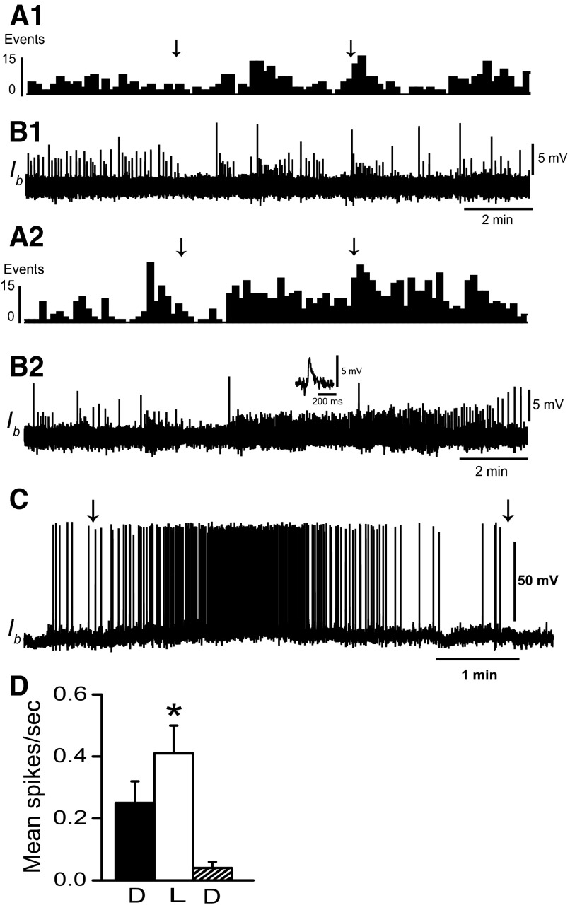
FIG. 3.
Light evokes EPSPs in type Ib interneurons. A1: histogram (10-s bin width) of EPSPs recorded from a dark-adapted type Ib interneuron, during 5 min of illumination of the photoreceptors and during 5 min in the dark following the offset of light. B1: intracellular recording of type Ib interneuron EPSPs contributing to the histogram in A1. The arrows above the histogram indicate the onset and offset of light-attenuated 1 log unit. A2: histogram of EPSPs recorded from the same dark-adapted type Ib interneuron as shown in A1 during 5 min of unattenuated light and 5 min in the dark following light offset. B2: EPSPs recorded from the type Ib interneuron contributing to the histogram in A2. The inset shows an example of an EPSP evoked by illumination of the photoreceptors. EPSP frequency increased as light intensity increased. Resting membrane potential of the Ib interneuron was 64–65 mV. C: light response of a type Ib interneuron depolarized to spike threshold prior to the onset of a 5-min period of illumination. D: group summary data (n = 5) of mean type Ib interneuron spike activity recorded during current depolarization in the dark (D), during 5 min of illumination of the photoreceptors (L) and during a 5-min period in the dark immediately following illumination (D). The arrows in C indicate the onset and offset of light that was attenuated 1 log unit; *P < 0.03.

