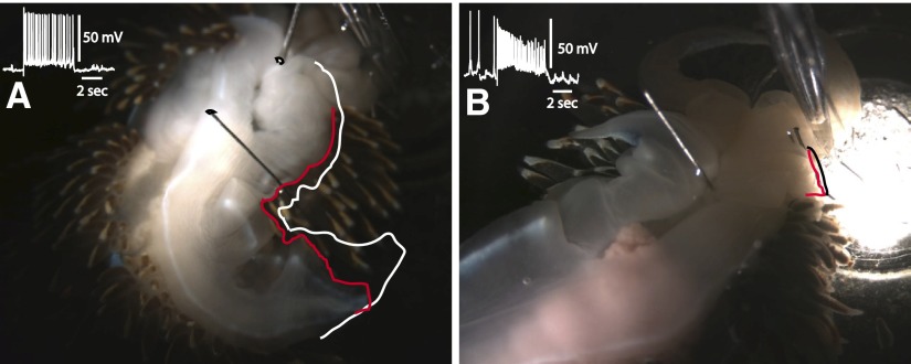
FIG. 7.
A: photograph of a representative semi-intact preparation before stimulation and at the termination of a 5-s depolarizing current pulse delivered to a type Ib interneuron. The white outline of the ipsilateral perimeter of the foot shows the foot immediately before stimulation and the red outline, the position of the foot at the end of the current pulse. Type Ib depolarization elicited a contraction and shortening of the rostral–caudal axis of the ipsilateral foot. The inset depicts spike activity of the Ib interneuron immediately before and during the 5-s current pulse. B: photograph of a representative semi-intact preparation before stimulation and at the termination of a 5-s depolarizing current pulse delivered to a type Is interneuron. The outline of the anterior foot before stimulation is shown in black and, at the end of the current pulse, the anterior foot position is indicated by the red line. The inset shows the spike activity of the Is interneuron immediately before and during the 5-s current pulse.

