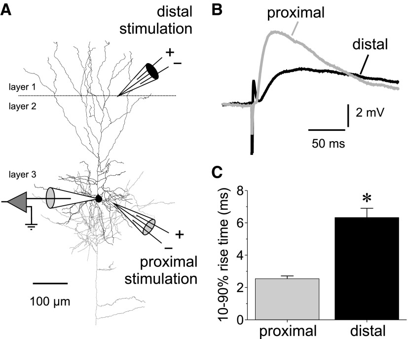
FIG. 2.
Stimulation of proximal and distal GABA synaptic inputs onto layer 3 pyramidal neurons. A: reconstruction of 1 of the cells recorded in this study showing the typical location of the stimulation electrodes. Distal stimulation was applied in near the layers 1/2 border. Proximal stimulation was applied 50–100 μm lateral to the soma of the recorded neuron. The pyramidal cell was reconstructed using Neurolucida (Microbrightfield, Williston VT), after staining to visualize the biocytin label was done as described previously (Gonzalez-Burgos et al. 2008). B: example average sweeps showing the differences in rising phase of IPSPs evoked in the same neuron by proximal vs. distal stimulation. C: summary graphs showing the statistically significant differences between the 10–90% rise time of IPSPs evoked by proximal and distal stimulation (rise time proximal IPSPs, 2.43 ± 0.21 ms, n = 30; distal IPSPs, 7.50 ± 0.87 ms, n = 24; independent samples t-test, t = 6.237, P < 0.00001).

