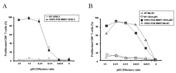
Figure 8.
CD8+ T cell clone activation by LV transduced pDC. In vitro antigen presentation capacities of LV transduced HLA-A2 pDC cells and Mo-DC. Cells were transduced with LV encoding the MART-1 peptide under the control of the PGK promoter. (A) Mature non-transduced (NT) and transduced (VSVG-PGK-MART-1) GEN2.2 or (B) CD34-pDC and Mo-DC were co-cultured with the HLA-A2 restricted CD8+ T-cell clone specific for the MART-1 peptide (LT12) stained with CFSE. After 5 days of co-culture, percentages of CD8+ dividing T-cells measured by flow cytometry were linearly correlated with the loss of CFSE fluorescence. The data in panel A are shown as the mean of triplicate and represent one out of 3 independent experiments whereas the data in panel B were performed once.

