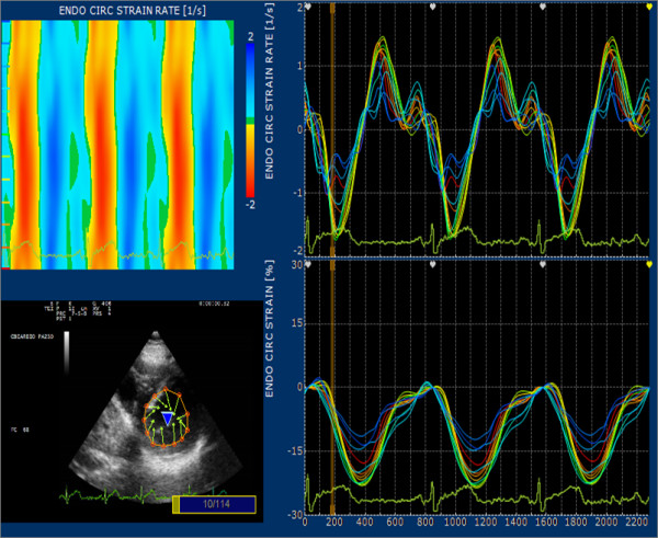
Figure 3.

Graph with parts of the processing result of a short axis view at mitral level. After selection of 12 equidistant point dividing the left ventricle in segments according to a 6 segments model; on left upper the system reports color analysis of circumferential SR and the curve report the values for circumferential Strain and SR for any point selected.
