Fig. 5.

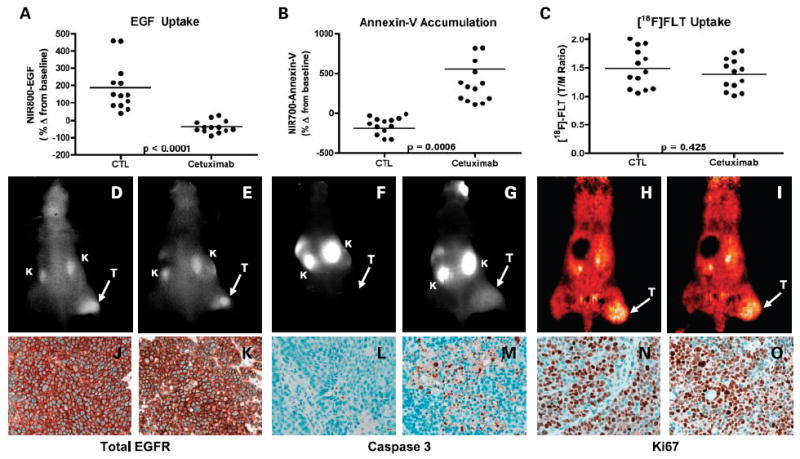
Noninvasive imaging assessment of response to EGFR blockade with cetuximab in DiFi xenograft-bearing mice. Treated and untreated cohorts bearing DiFi xenograft tumors were simultaneously imaged with NIR800-EGF, NIR700-Annexin V, and [18F] FLT PET. Following cetuximab treatment, DiFi tumors exhibited significantly reduced NIR800-EGF uptake (A) and increased NIR700-Annexin V uptake (B) compared with untreated controls. No statistical difference in [18F] FLTuptake was observed between treated and untreated mice. Units are defined as tumor (T)/muscle (M) Ratio (C). Representative NIR800-EGF, NIR700-Annexin V, and [18F] FLT PET images collected from an individual control (D, F, and H) and treated (E, G, and I) mouse. Strong agreement between the imaging metrics of response and standard immunohistochemistry was observed. Tumors from control (J) and treated (K) animals exhibited similar levels of total EGFR. Treated animals (M) exhibited elevated caspase-3 staining compared with untreated cohorts (L). No discernible difference in Ki-67 staining was observed between tumors from control (N) and treated cohorts (O).
