
Figure 2.
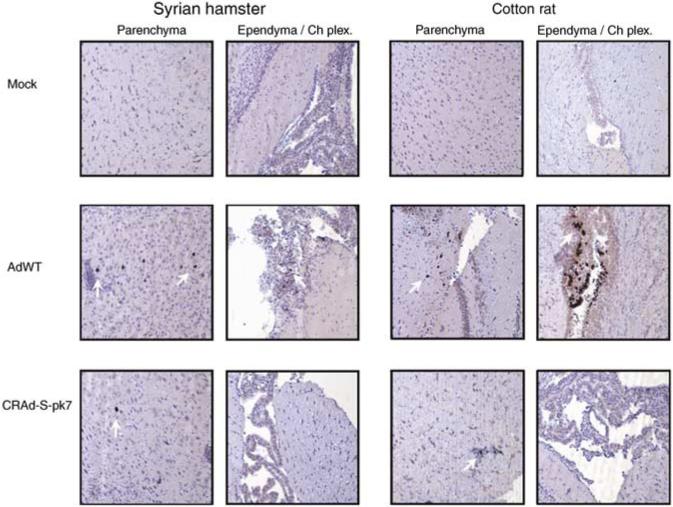
Immunohistochemical staining for viral antigen E1A in Syrian hamster (SH) and cotton rat brain slices. At day 7 after i.c. injection of CRAd-S-pk7 or AdWT, SHs and cotton rats were killed and brain samples were fixed, sliced and analyzed for the presence of viral antigen E1A by immunohistochemistry. White arrows point to cells that stained positive for E1A antigen. Representative × 10 power fields are presented from brain parenchyma and ependyma/choroid plexus from SHs (left) and cotton rats (right). Mock-treated animals (top) did not exhibit any staining, whereas animals injected with AdWT (middle) or CRAd-S-pk7 (bottom) stained positively for E1A in some cells.