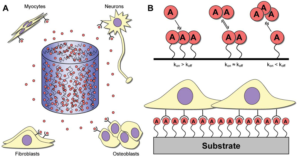
Fig. 1. Release mechanisms for protein and DNA.
(A) Inductive factors (red circles) may be embedded or encapsulated within hydrogels or microspheres, which may, in turn, be used to form 3-D scaffolds that are capable of releasing factors at the site of implantation. The highest factor concentration exists within the scaffold, with lower concentrations found within the surrounding tissue. Delivered factors target a variety of different cell populations (e.g., myocytes, neurons, fibroblasts or osteoblasts) for various applications (e.g., muscle, nerve, bone, wound healing). (B) Substrate immobilization is characterized by factors being bound to a biomaterial. The interaction between the factor and biomaterial can be (top): (1) kon ≫ koff, such that the factor is effectively bound irreversibly; (2) kon ≈ koff, where the factor associates and dissociates from the surface at roughly equal rates; and (3) kon ≪ koff, such that the factor is loaded onto the biomaterial and dissociates to serve as a delivery vehicle. Cells interact with the immobilized factors upon interaction with the biomaterial (bottom).

