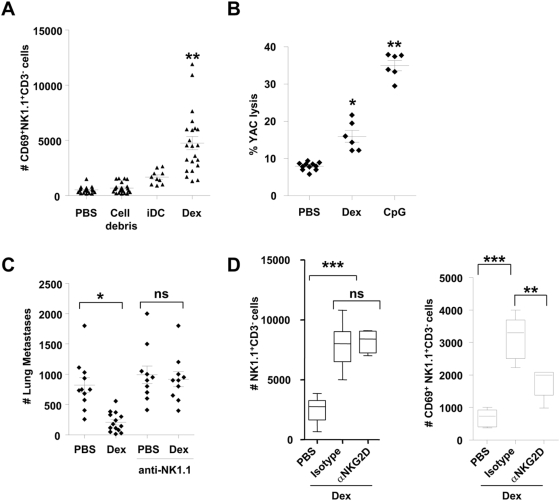
Figure 2. Mouse Dex promoted NK cell activation: a role for NKG2D.
A. Dex induced NK cell activation in the draining lymph node. Absolute numbers of CD3− NK1.1+ CD69+ cells in the draining lymph node following inoculation of 10 µg of mouse Dex, or 3×10⁵ iDC or 10 µg of irrelevant pelleted proteins (cell debris) or PBS. Each dot represents the result in one mouse. B. Dex stimulated splenic NK cytotoxicity. Killing assays on splenocytes against YAC-1 targets at 200∶1 and 50∶1 (not shown) after intradermal inoculations of 10 µg of Dex every other week for 8 weeks and sacrifice 48 hrs after the last immunization, or 24 hrs after a single subcutaneous injection of 10 µg of CpG ODN. C. Therapy with unpulsed Dex reduced number of metastases. Intradermal inoculation of 20 µg of exosomal proteins or PBS 5 days after intravenous injection of 3.105 B16F10 tumor cells. Depletion of NK cells was achieved using 3 administrations of anti-NK1.1 mAbs or isotype control mAbs. Mice were sacrificed on day 10 for enumeration of lung metastases. D. A role for NKG2D in Dex-mediated NK cell triggering. Enumeration of CD3−NK1.1+ cells (left panel) and CD3− NK1.1+CD69+ cells (right panel) in the draining lymph node following intradermal inoculation of PBS or 10 µg of mouse Dex in the presence of anti-NKG2D blocking mAb (αNKG2D) or control isotype mAb. The graphs depict the data of 4 pooled experiments (2 for B.). Means and SEM are shown. * p<0.05, ** p<0.01, *** p<0.001 and ns: “non significant”.

