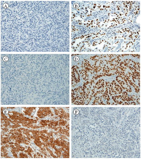
Figure 3.
Medullary carcinoma showing negative staining with MLH-1 (A) and poorly differentiated carcinoma showing positive nuclear staining with MLH-1 (B). Medullary carcinoma showing negative staining with CDX2 (C) and poorly differentiated carcinoma showing positive nuclear staining with CDX2 (D). Medullary carcinoma showing positive nuclear and cytoplasmic staining with calretinin (E), and poorly differentiated carcinoma showing negative staining for calretinin (F). (x50 magnification)

