Figure 1.

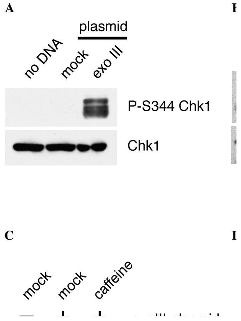
Exonuclease III-treated plasmid DNA can induce phosphorylation of checkpoint proteins. (A) ExoIII-treated DNA induces Chk1 phosphorylation at S344. Mock- or exoIII-treated plasmid (2.5 ng) was incubated with 5 μL Xenopus NPE for 30 minutes. 1 μL of each sample was run on an 8% SDS-PAGE gel and analyzed for phospho-Chk1 (P-S344) and total Chk1 by immunoblotting. (B) ExoIII-treated DNA induces RHR complex phosphorylation. ExoIII-treated plasmid DNA (5 ng) was incubated in 10 μL HSE for 30 minutes. 1 μL in vitro translated RHR complex (35S-Rad9, 35S-Hus1, cold Rad1) was added to the extract prior to plasmid addition to trace Rad9 and Hus1 phosphorylation. 1 μL extract was run out on a 10% SDS-PAGE gel. Rad9 and Hus1 were visualized by autoradiography, and Rad1 was analyzed by immunoblotting with anti-Rad1 antibodies. (C) Rad1 is phosphorylated on T5. ExoIII-treated plasmid DNA (2.5 ng) was incubated in 5 μL HSE for 30 minutes. To inhibit ATR activity, 5 mM caffeine was added to the extract prior to the addition of the plasmid. Rad1 was analyzed by immunoblotting with anti-Rad1 and anti-P-T5 phospho-specific antibodies. (D) ExoIII treatment of a nicked plasmid creates a heterogenous set of structures. Plasmid DNA (pEYFP) containing nicks was mock-treated or treated with 100 U exonuclease III for 2′ at 37 degrees. The mock- and exoIII-treated plasmid was electrophoresed along with an equivalent amount of single-stranded M13 DNA on an 0.8% agarose gel and stained with ethidium bromide. [Figures 1A and 1C reprinted with permission from Molecular Biology of the Cell, copyright 2006.]
