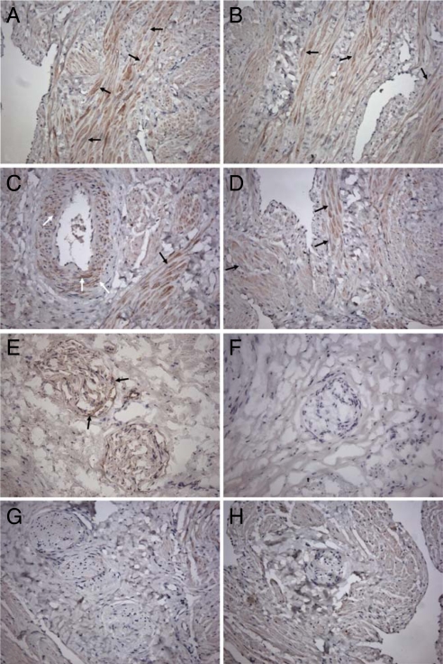
Fig. 2.
Immunochemistry for CBS and CSE in HCC. (A–D) Immunohistochemical detection of CSE and CBS in HCC tissue. Immunoreactivity and nuclear staining appear brown (DAB) and blue (hematoxylin counterstain), respectively. CSE was detected in trabecular muscular tissue (A and B, black arrows) and vascular smooth-muscle cells (C, white arrows). Immunoreactivity for CBS was mostly observed in trabecular muscular tissue (D, black arrows). Results illustrated are from a single experiment and are representative of 3 different specimens. (Original magnification, 200×.) (E–H) Immunohistochemical detection of CSE and CBS in HCC nerve fibers. Immunoreactivity and nuclear staining appear brown (DAB) and blue (hematoxylin counterstain), respectively. CSE was detected in nerve fibers in cryostat (E, arrows) and not in paraffin (G) sections. Both cryostat (F) and paraffin (H) sections lacked immunoreactivity for CBS. Results illustrated are from a single experiment and are representative of 3 different specimens. (Original magnification, 200×.)

