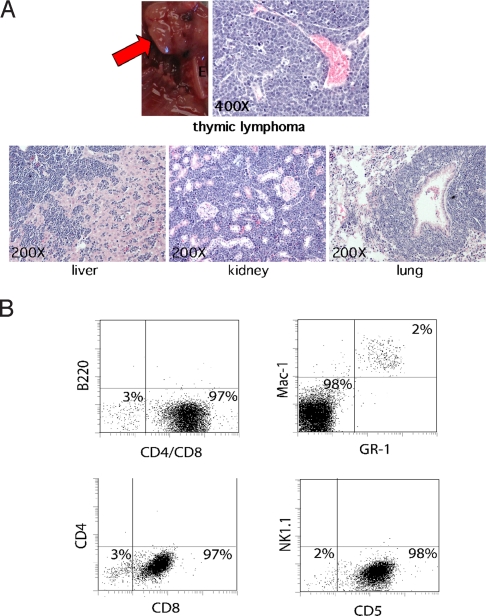
Fig. 5.
High-grade thymic T cell lymphomas induced in ASPP2+/− mice after γ-irradiation. (A) Thymic lymphoma (red arrow) in an ASPP2+/− mouse. H&E-stained microscopic sections of lymphoma-infiltrated liver, kidney, and lung from this mouse. (B) Immunophenotyping of tumor cells in unfractionated bone marrow from this mouse. A total of 2% of normal myeloid progenitors coexpressed Mac-1 and Gr-1 (Upper Right). A total of 98% of the bone marrow was replaced by tumor cells expressing CD5 and CD8 (Lower), but not B220 (Upper Left), CD4, and Nk1.1 markers (Lower).

