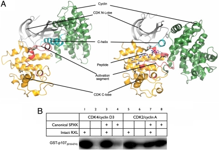
Fig. 1.
Structure and substrate selection in CDK4/cyclin D3. (A) The fold of CDK4/cyclin D3 (left side) is compared with that of CDK2/cyclin A in complex with a substrate peptide (PDB ID code 1QMZ; right side). Important structural and regulatory elements referred to in the text are indicated on each structure and correspond to the CDK N-lobe (light gray), the C-helix (cyan), the CDK C-lobe (yellow), the activation segment (red), and the cyclin subunit (green). (B) The activity of CDK4/cyclin D3 toward pRb family members highly depends on substrate recruitment. See Results for details.

