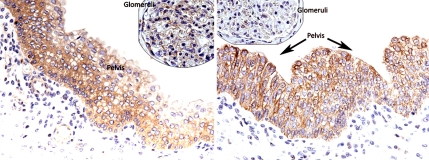
Fig. 3.
IHC staining for ARHGEF6 and STMN3 on control kidney tissue. Cytoplasmic staining is observed in the pelvic urothelium with ARHGEF6 and STMN3. Glomerular staining is also observed for ARHGEF6. (Left) ARHGEF6 shows positive staining in renal pelvis and glomerulus. Faint staining is seen in proximal tubules, podocytes and epithelial cells. (Right) STMN3 shows positive staining exclusively in the pelvis. Mild staining was seen in proximal tubules, podocytes, and epithelial cells.

