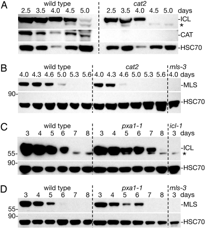
Fig. 2.
Mutants varying peroxisome H2O2 production exhibit altered ICL and MLS degradation kinetics. (A and B) Immunoblots of protein extracts from wild-type and cat2 cotyledons (12 per lane) were probed with α-ICL, α-catalase (CAT), α-HSC70, and α-MLS antibodies. (C and D) Immunoblots of total protein extracts from wild-type and pxa1–1 cotyledons (12 per lane) were sequentially probed with α-ICL and α-HSC70 (C) or α-MLS and α-HSC70 (D) antibodies. Null mls-3 and icl-1 (8) mutants provide controls for the MLS (B, D) and ICL (C) antibodies, respectively. Asterisk marks a cross-reacting band detected by the α-ICL antibodies that remains present in the icl-1 null mutant, and positions of molecular weight markers (in kDa) are indicated on the left.

