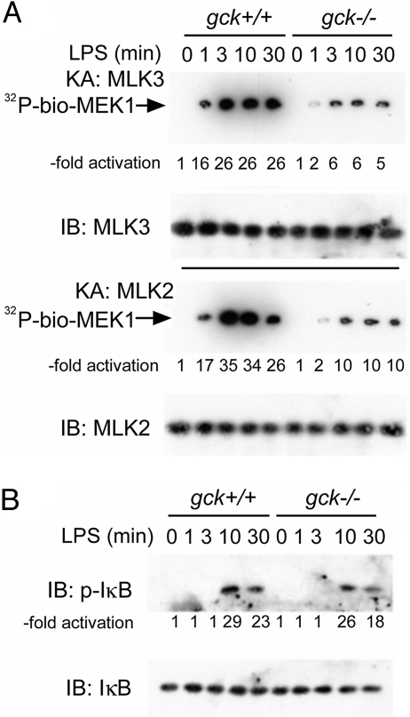
Fig. 2.
Disruption of gck impairs LPS activation of macrophage MLKs-2 and -3, but not NF-κB. BMMs are prepared from gck+/+ and −/− mice and treated with LPS for the indicated times. (A) MLK2 or MLK3 is immunoprecipitated, as indicated, from crude cell extracts and assayed by using biotinylated-MEK1 and γ[32]ATP as substrates. The −fold activation is shown. (B) Extracts are prepared from BMMs stimulated as above and subjected to SDS/PAGE and immunoblotting with the indicated antibodies. Levels of phospho-IκB are quantitated by ImageJ. IB: immunoblot; KA: kinase assay.

